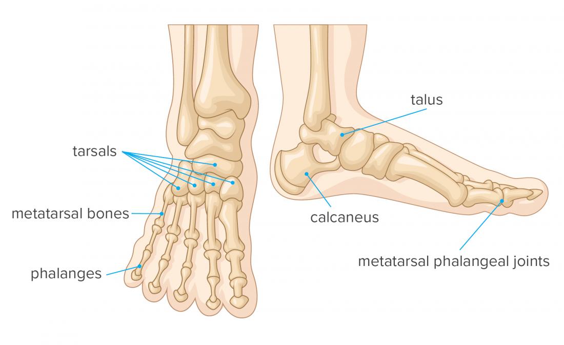
Foot Bone Structure Diagram. You can see the toes on the top and the heel on the bottom while the arch and sole of the foot are made up of a thick web of ligaments holding the bones together. There are also 2 sesamoid bones not shown located under the 1st MTP joint.

You can see the toes on the top and the heel on the bottom while the arch and sole of the foot are made up of a thick web of ligaments holding the bones together. Understanding the structure of the foot is best done by looking at a foot diagram where the anatomy has been labeled. The midfoot is a pyramid-like collection of bones that form the arches of the feet.
The foot begins at the lower end of the tibia.
Bones muscles tendons and nerves which will each give slightly different foot pain symptoms. Together the tarsals form the arch of the foot. Notice that the great toe only has a proximal and distal phalange. Proximal intermediate and distal.

