Occipital Bone Diagram. Bone Diagram Forehead Frontal bone Nose bones Nasals Cheek bone Zygoma Upper jaw Maxilla Lower jaw Mandible Breast bone Sternum Upper arm bone Humerus Lower arm bone. The occipital bone of the pig is flattened and elongated that forms the caudal part of the skull.
The occipital bone of dog skull. The four regions that are integral to the occipital bone anatomy include. A newborn has a frontal bone that consists of two parts separated by the frontal suture.
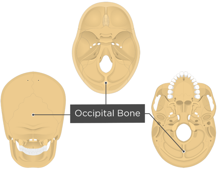
These four regions surround the foramen magnum which is a large hole that is located at the center of the occipital bone.
Inner surface of Occipital bone Temporal bone. Sutures connect cranial bones and facial bones of the skull. The muscle has a frontal belly and an occipital near the occipital bone on the posterior part of. Forms the posterior part of the skull and the base of the cranium.
