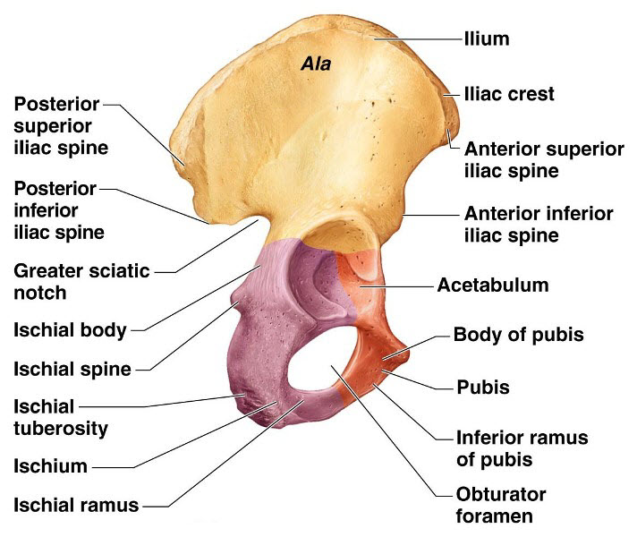
Hip Bone Anatomy Diagram. The hip bone is comprised of the three parts. The hip joint is a ball-and-socket synovial joint formed between the os coxa hip bone and the femur.
Hip Bone Anatomy. Ad Non-Invasive Osteoarthritis Treatments. It is the largest ball-and-socket joint in your body.
The thigh bone or femur and the pelvis which is made up of three bones called ilium ischium and pubis.
Normal hip joint and bone with skeletal disorder that characterized by low bone mass. Difference and comparison Bone medical health questions and osteoporosis illustration concept as a close up diagram of the inside of human skeletal hip bones with a magnification glass showing a normal healthy condition degrading to abnormal. The hip itself is a ball and socket joint much like the shoulder. The hip joint is the largest ball and socket joint in the body.
