Diagram Of Skeletal Muscle. The skeletal muscle fibers are crossed with a regular pattern of. Skeletal muscle is composed of bundles of myofibrils or muscle cells that are long and fibrous in appearance.
Skeletal muscle diagram 308. Skeletal muscle is one of the three types of muscles in the human body- the others being visceral and cardiac muscles. Within each muscle fiber are myofibrils long cylindrical structures that lie parallel to the muscle fiberMyofibrils run the entire length of the muscle fiber.
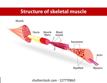
Diagram of skeletal muscle fiber structure.
Each skeletal muscle has three layers of connective tissue that enclose it provide structure to the muscle and compartmentalize the muscle fibers within the muscle Figure 1021. The skeletal system consists of 206 bones that make up the internal framework of the body called the skeleton. If myosin molecules are unable to bind to other myosin molecules this prevents muscle contraction. There is a printable worksheet available for download here so you can take the quiz with pen and paper.
