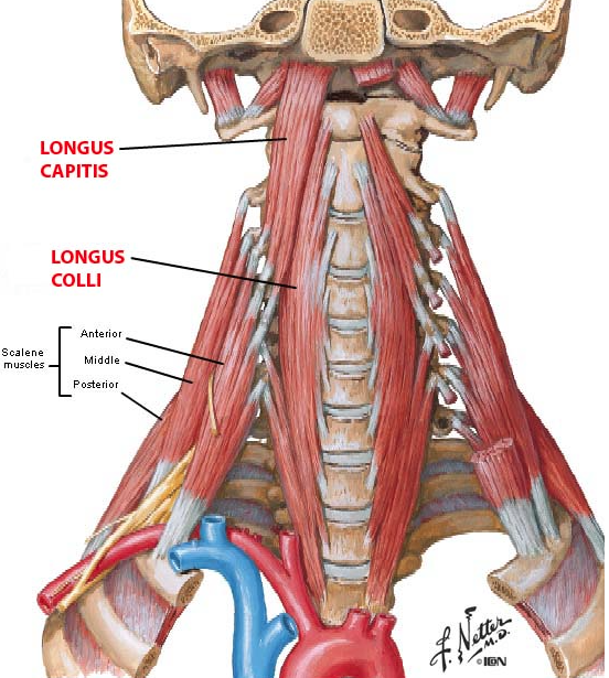
Cervical Muscle Anatomy. The largest muscles of the posterior cervical region are the sternocleidomastoid and the trapezius. As viewed from the side the cervical spine forms a lordotic curve by gently curving toward the front of the body and then back.

Majority of flexionextension of the neck and lateral bending occur here. When contracted the scalenes cause the first and second ribs to elevate and the neck to bend to the side. The erector spinae are a group of many muscles that attach along the back of the spine.
Maximal lateral bending occurs.
The sternocleidomastoid muscle originates at the anterior surface of the manubrium and the medial one-third of the clavicle. When contracted the scalenes cause the first and second ribs to elevate and the neck to bend to the side. The Superficial Fascia of the neck is a thin lamina investing the Platysma and is hardly demonstrable as a separate membrane. The cervical rhomboid muscle Musculus rhomboideus cervicis is the cervical part of the rhomboid muscle which originates form the spinous processes of the the cervical vertebrae.
