Cardiac Muscle Cell Diagram. The extracellular fluid the SR and the cytoplasm. A membrane potential is the difference in electrical potential between the interior and the exterior of the cell membrane.
Cardiac Muscle And Electrical Activity Anatomy And Physiology Ii from courses.lumenlearning.com
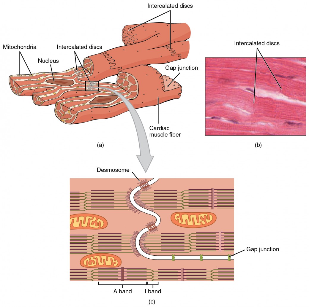
Cardiac muscle cells usually have a single central nucleus. The fibrils do not run strictly parallel to each other but rather branch in a complex pattern. Cardiac muscle fibers cells also are extensively branched and are connected to one another at their ends by intercalated discs.
NOTES NOTES MUSCLES MUSCULAR SYSTEM ANATOMY.
Cardiac Cycle Diagram The diagram below represents the different phases of the cardiac cycle. Cardiac muscle fibers also possess many mitochondria and myoglobin as ATP is produced primarily through aerobic metabolism. NOTES NOTES MUSCLES MUSCULAR SYSTEM ANATOMY. The atrial systole ventricular diastole ventricular systole and ventricular diastole are clearly mentioned in the cardiac cycle diagram given below.